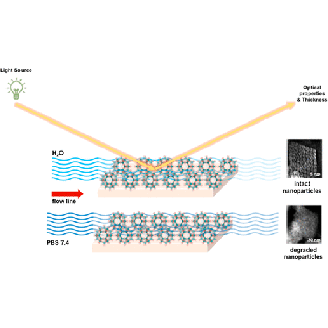
Porous iron (III) carboxylate metal−organic frame-works (MIL-100; MIL stands for Material of Institute Lavoisier) of submicronic size (nanoMOFs) have attracted a growing interest in the field of drug delivery due to their high drug payloads, excellent entrapment efficiencies, biodegradable character, and poor toxicity. However, only a few studies have dealt with the nanoMOF degradation mechanism, which is key to their biological applications. Complementary methods have been used here to investigate the degradation mechanism of Fe-based nanoMOFs under neutral or acidic conditions and in the presence of albumin. High-resolution STEM-HAADF coupled with energy-dispersive X-ray spectroscopy enabled the monitoring of the crystalline organization and elemental distribution during degradation. NanoMOFs were also deposited onto silicon substrates by dip-coating, forming stable thin films of high optical quality. The mean film thickness and structural changes were further monitored by IR ellipsometry, approaching the “sink conditions” occurring in vivo. This approach is essential for the successful design of biocompatible nano-vectors under extreme diluted conditions. It was revealed that while the presence of a protein coating layer did not impede the degradation process, the pH of the medium in contact with the nanoMOFs played a major role. The degradation of nanoMOFs occurred to a larger extent under neutral conditions, rapidly and homogeneously within the crystalline matrices, and was associated with the departure of their constitutive organic ligand. Remarkably, the nanoMOFs’ particles maintained their global morphology during degradation.
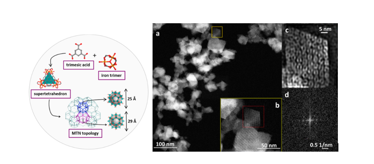
Schematic representation of the structure of MIL-100(Fe) obtained by the self-assembly of BTC and iron, forming super tetrahedra and then 3D mesoporous cages of 25 and 29 Å, accessible through microporous pentagonal windows of around 5 Å and microporous hexagonal windows of 8.6 Å. Typical STEM-HAADF images of synthesized nanoMOFs (RT). (a) Unprocessed image; (b) magnified image; (c) inverse fast Fourier transform (FFT) filtered HAADF-STEM image, and (d) typical FFT pattern which reveals the crystallinity of the particles.
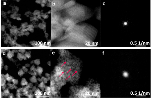
STEM-HAADF images of nanoMOFs after 48 h of degradation in PBS (Phosphate Buffered Saline) at pH 5.4 and (d,e) in PBS at pH 7.4. The concentration was 0.25 mg mL−1.
Red arrows show the appearance of large pores (red arrows) indicating degradation. (c,f) FFT patterns of degraded nanoMOFs indicate the loss of crystallinity.
Reference
Advanced Characterization Methodology to Unravel the Biodegradability of Metal–Organic Framework Nanoparticles in Extremely Diluted Conditions
I. Christodoulou1, G. Patriarche2, C. Serre3, C. Boissiere4, R. Gref1
ACS Applied Materials & Interfaces, 16 (2024) 13353-14384
https://pubs.acs.org/action/showCitFormats?doi=10.1021/acsami.3c18958&ref=pdf
Affiliations
1 Institut des Sciences Moléculaires d’Orsay (ISMO), Université Paris-Saclay, CNRS UMR 8214, 91405 Orsay, France
2 Centre de Nanosciences et de Nanotechnologies (C2N), Université Paris-Saclay, CNRS UMR 9001, 91120 Palaiseau, France
3 Institut des Matériaux Poreux de Paris, Ecole Normale Supérieure, ESPCI Paris, CNRS, PSL University, 75005 Paris, France
4 Laboratoire de Chimie de la Matière Condensée de Paris (LCMCP), Sorbonne Université, CNRS, Collège de France, 75005 Paris, France