
News from the Institute of Molecular Sciences of Orsay - July 25, 2018 (released in French)
Researchers from ISMO and C2N publish the first comprehensive study of the effects and artifacts that form the image in optical microscopy of the Fourier plane, on periodic samples. These artifacts, used to exceed the diffraction limit, can be exploited, for example, for nano-positioning and autofocus applications in light microscopy. This work is published in the Journal of Applied Physics and the article is briefly featured on the AIP Scilight website.
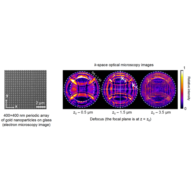
k-space optical microscopy (or Fourier-plane imaging) is a technique increasingly used in all fields of photonics including biosensing, photonic crystals, plasmonics, and single-molecule studies. It consists in the imaging of the back focal plane of a microscope objective ("k" refers to the coordinates in Fourier space). However, the performance of this technique under various illumination conditions, as well as its artifacts and the ways to overcome or even benefit from these artifacts, have rarely been addressed in the literature.
In this article, a group from the Institute of Molecular Sciences of Orsay - ISMO (CNRS/UPSUD) and a group from the Center for Nanoscience and Nanotechnology – C2N (CNRS/UPSUD) jointly provided a detailed description of the performance and inherent artifacts of k-space optical microscopy for the study of periodic nanoparticle arrays. They used the various illumination configurations available on an inverted optical microscope, i.e., with transmitted or reflected, collimated or focused, coherent or incoherent light. The aim of this work is to help readers exploit the full potential of this technique and to foster the development of innovative applications.
Among the most interesting results are images of k-space optical microscopy of periodic samples illuminated in reflection with a focused laser beam. These images result from the interference of the reflected and diffracted light beams. Crucial information contained in the phase of the field, which can be related either to the geometry of the sample or to the relative position of the focal point and the sample, are thus converted into intensity variations. This can be used for the accurate control of the sample position in optical microscopy, with a precision well beyond the diffraction limit of light. Possible technological spin-offs of these results include applications to autofocus and repositioning in optical devices, e.g., in writing and readout operations for optical data storage.
Source : “k-space optical microscopy of nanoparticle arrays: opportunities and artifacts,” Jean-François Bryche, Grégory Barbillon, Bernard Bartenlian, Gérald Dujardin, Elizabeth Boer-Duchemin, Eric Le Moal, Journal of Applied Physics (2018).
Contact :
- Eric Le Moal, Institut des Sciences Moléculaires d’Orsay (CNRS - Université Paris-Sud)
- Bernard Bartenlian, CNRS researcher at C2N
- Jean-François Bryche, PhD student at C2N